


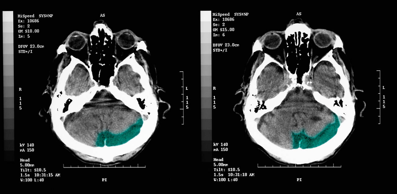
Alzheimer’s disease may begin altering the brain structure as much as ten years before a person notices memory problems, according to a new study from Johns Hopkins University. Scientists identified two specific areas, the entorhinal cortex and the amygdala, that show signs of deterioration earlier than the hippocampus, which has traditionally been the primary focus for detecting the disease.
The discovery suggests that doctors might be able to recognize the condition long before symptoms develop. Starting treatment during this silent phase could potentially protect the brain while it still has significant function remaining.
Early Tissue Loss in Memory and Emotion Centers
The researchers analyzed hundreds of brain scans from two independent studies: the Alzheimer’s Disease Neuroimaging Initiative and the Biomarkers of Cognitive Decline Among Normal Individuals study. These scans tracked individuals across different stages of the disease, including those with no symptoms, those with cognitive impairment, and patients who later died from the condition.

In every group, the team observed a consistent pattern. The entorhinal cortex and amygdala, located near the hippocampus, experienced greater relative tissue loss during the early stages of Alzheimer’s than the hippocampus itself. The entorhinal cortex is responsible for retrieving stored memories, while the amygdala processes emotional memories.
Michael Miller, a biomedical engineer and senior author of the study, emphasized the timeline of these changes. He noted that the brain undergoes a five to ten year period of alteration before clinical symptoms appear. Treating the disease during the two to four year mark, rather than after fifteen years of decline, represents the main promise of this research.
Connection to Tau Protein Accumulation
The study also linked the observed tissue loss to tau, a protein closely associated with Alzheimer’s. Under normal conditions, tau helps maintain the internal structure of nerve cells. In Alzheimer’s, however, tau accumulates abnormally and forms tangles that disrupt healthy brain function.
Using positron emission tomography (PET) imaging and digital pathology, the scientists found that areas of gray matter shrinkage overlapped with regions showing tau-related changes. Gray matter contains large numbers of nerve cell bodies and is essential for processing information throughout the brain. This overlap strengthens the possibility that changes in the entorhinal cortex and amygdala could serve as useful indicators of early Alzheimer’s disease.
Marilyn Albert, co-author and director of the Johns Hopkins Alzheimer’s Disease Research Center, stated that the goal is to treat individuals before symptoms develop so that the progression can be delayed or stopped. Meaghan Morris, a neuropathologist and co-author, added that knowing who will experience cognitive decline is crucial for better risk stratification and earlier treatment.
The findings were published in NeuroImage Reports and presented at Neuroscience 2026, the annual meeting of the Society for Neuroscience. While the results are promising, the team notes that more research is needed before these brain changes can be used as a reliable diagnostic tool. The next step involves investigating molecular changes surrounding abnormal tau accumulation to identify new targets for therapies.
Source: ScienceDaily
