


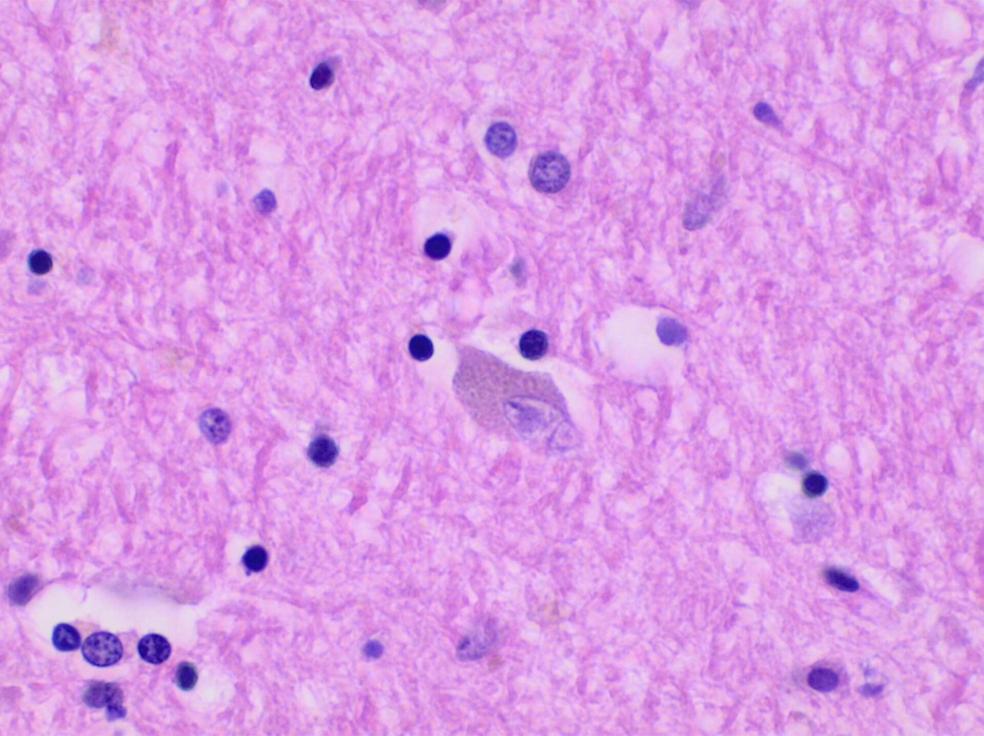
Researchers from the University of Pennsylvania have presented findings that offer rare direct evidence from a human brain showing how removing amyloid plaques may protect against other forms of brain damage. The study, published in JAMA and presented at the 2026 Alzheimer’s Association International Conference, examined the brain of a man who received treatment for Alzheimer’s disease and later donated his tissue for scientific research.
Uneven Clearance in the Same Brain
The investigation focused on a patient in his fifties who was diagnosed with mild cognitive impairment. He participated in a clinical trial for aducanumab, receiving 30 doses of the medication over four and a half years. He died approximately four years after his final dose, more than a decade after his first symptoms appeared.
When pathologists examined the donated brain, they found a striking contrast between neighboring regions. The raised ridges of the brain’s surface, known as gyri, had lost most of their amyloid plaques. These same areas contained very little accumulated tau, a protein that forms tangles inside neurons and is strongly linked to cell death.
In contrast, the grooves between these ridges, called sulci, still held considerable amounts of amyloid. These regions exhibited substantially higher levels of tau tangles, inflammation, and tissue degeneration. This patchwork distribution matched earlier scans taken while the patient was alive, which had already indicated that the drug’s effect varied across different parts of the brain.
“This case represented a unique scenario in which some regions cleared amyloid and others did not,” said co-senior author Edward B. Lee, MD, PhD. “It allowed us to directly compare what happened next in neighboring brain regions and better understand the relationship between amyloid, tau, and neurodegeneration.”
Implications for Current Treatments
The findings suggest that clearing amyloid thoroughly, particularly early in the disease, could help limit the accumulation of tau and slow the progression of brain damage. Beta-amyloid collects outside nerve cells to form sticky deposits, while tau accumulates inside neurons. Both processes contribute to the progressive damage that causes memory loss and cognitive decline.
Treatments designed to eliminate amyloid have changed how Alzheimer’s is managed in recent years. While aducanumab received accelerated approval before being withdrawn from the market, newer medications like donanemab (Kisunla) and lecanemab (Leqembi) have received full FDA approval. These drugs target amyloid through similar mechanisms to the therapy received by the patient in this study.
David Wolk, MD, co-director of the Penn Memory Center, noted that seeing both disease patterns side-by-side provided a rare opportunity to understand how amyloid removal affects other proteins. He stated that the findings provide some of the clearest human evidence to date that anti-amyloid therapies may limit the accumulation of tau and slow the brain changes that lead to memory loss.
Although the study involved only one individual, the contrast between different brain regions suggests that substantial amyloid removal may influence other disease processes long after treatment has stopped. The results also raise questions about how the benefits of Alzheimer’s drugs are measured, as clinical trials commonly evaluate patients over approximately 18 months, while some protective effects might take longer to become apparent.
Researchers now hope to understand why certain parts of the brain respond more effectively to amyloid-clearing treatments than others. Identifying the biological mechanisms responsible for plaque removal could lead to better therapies. Another major question is whether beginning treatment before extensive tau tangles develop could substantially delay the disease or even prevent it from emerging.
Source: ScienceDaily
