


Researchers at the RIKEN Pioneering Research Institute in Japan have created a method that forces cancer cells to manufacture their own destruction. Led by Katsunori Tanaka, the team published their findings in Nature Communications, describing a process where malignant cells use excess carbon monoxide to synthesize toxic compounds directly within their own boundaries.
Traditional chemotherapy often damages healthy tissue because toxic drugs circulate throughout the entire body. This new strategy aims to eliminate that collateral damage. The team designed a system where relatively harmless compounds enter the body and only become active when they encounter the specific chemical environment of a tumor.
Using Tumor Chemistry as a Trigger
Cancer cells naturally produce higher levels of carbon monoxide than normal cells. While this gas is often associated with combustion engines, it is also a byproduct of cellular metabolism in tumors. The researchers identified this excess gas as a potential trigger for a chemical reaction that would not occur in healthy tissue.
The core of the system relies on a metal catalyst known as a palladacycle. When this catalyst reacts with carbon monoxide, it produces an organic molecule called phenanthridinone. Certain versions of this molecule are already known to be toxic to cells and are used in existing anticancer medications. The goal was to make the body produce these toxic agents only where the carbon monoxide levels are high enough to drive the reaction.
A significant hurdle in using metal catalysts inside living organisms is that cells contain substances like glutathione, which quickly deactivate the catalyst. To solve this, the team enclosed the palladacycle within albumin, a common blood protein. This protein shell protected the catalyst from being neutralized by cellular defenses, allowing it to reach its target intact.
Selective Activation in Cancer Lines
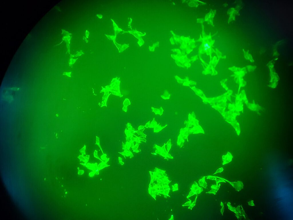
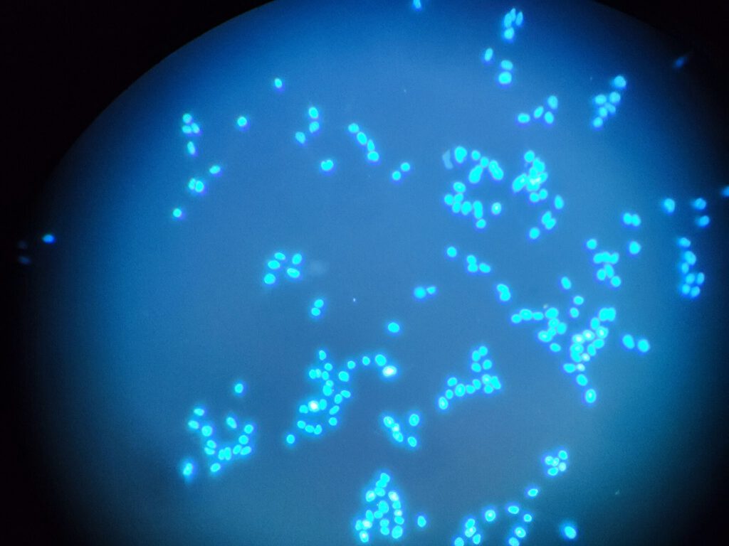
The team tested the albumin-encased catalyst on cultured human liver, cervical, breast, and lung cancer cells. They also exposed noncancerous cell lines to the same treatment to check for unintended reactions. After 48 hours, phenanthridinone was detected in the cancer cells but remained absent in the healthy cells.
This selectivity confirmed that the system worked as intended. The catalyst remained inactive in normal cells due to insufficient carbon monoxide, while it successfully triggered drug production in malignant cells. The researchers further modified the system to ensure the newly formed toxic compound could detach from the albumin carrier and move freely within the cell to cause damage.
Safety concerns regarding the bloodstream were also addressed. Carbon monoxide is present in the blood, but it is mostly bound to hemoglobin. Laboratory tests showed that this bound gas did not trigger the reaction, preventing the premature creation of toxic drugs in the circulatory system. This suggests the compound can travel through the body without harming blood cells.
Tanaka described the method as a significant step in therapeutic in vivo synthetic chemistry. The approach uses molecules naturally found in the body, such as disease-associated metabolites, as feedstocks to create therapeutic agents directly at the site of the disease.
The next phase of the research involves moving from cell cultures to animal models. If these tests confirm the safety and efficacy of the method, it could lead to a new class of cancer treatments that rely on the tumor’s own chemistry to fight itself.
Source: ScienceDaily
