


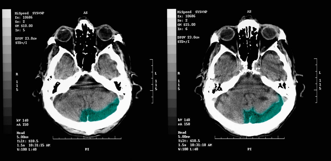
Walking into a crowded parking lot and forgetting where you parked your car is a common frustration, but for older adults, this lapse in episodic memory can signal deeper neurological changes. A new study from the University of California, Berkeley, identifies a specific pattern of brain activity during sleep that correlates with memory decline and the accumulation of tau protein, a key marker for Alzheimer’s disease.
Neuroscientist Omer Sharon and his colleagues found that as tau builds up in the frontal cortex, the brain’s ability to generate coordinated electrical waves during deep sleep deteriorates. These waves, which normally travel across large regions of the brain to consolidate memories, become fragmented and isolated. The researchers refer to these disconnected patterns as “lonely waves.”
Disrupted Sleep Patterns in Aging Brains
During non-REM sleep, large groups of neurons switch off and back on in a synchronized sequence. This coordinated activity produces slow electrical waves that typically originate in the frontal cortex and cascade across the brain. This process is critical for transferring short-term experiences into long-term storage.
The study compared electroencephalogram (EEG) data from cognitively healthy young adults in their early 20s with older participants in their mid-60s to mid-70s. In younger participants, slow waves moved together across distances roughly equivalent to the width of a hand on the scalp. In older adults, these waves traveled shorter distances and appeared more independently, lacking the coordinated grouping seen in younger brains.
“This is the first time we’ve shown this relationship between tau and how it messes with memory consolidation by attenuating traveling slow waves that originate in the frontal cortex,” Sharon said. “Our study shows that for memory, it’s critical that these events cascade in sequence across large parts of the brain.”
Tau Accumulation and Cognitive Decline
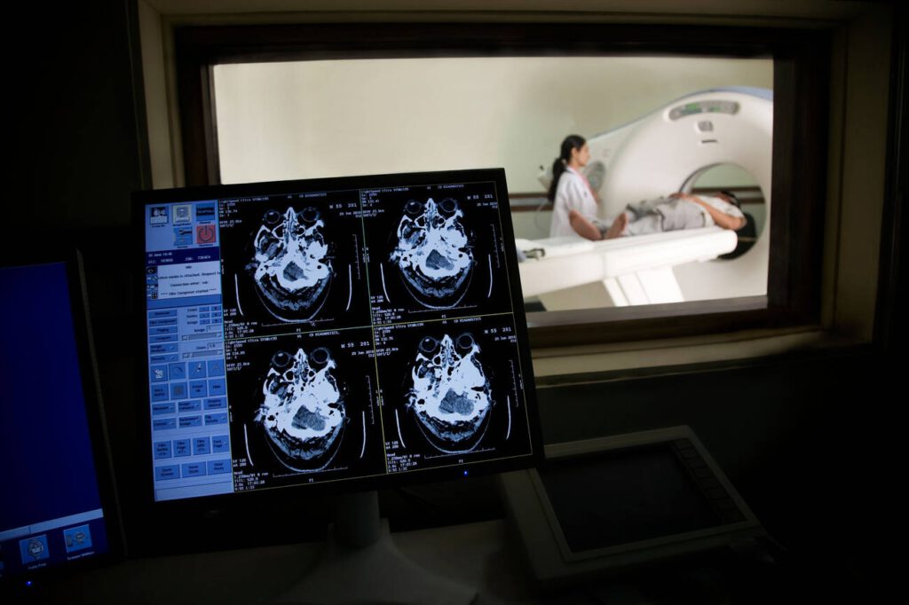
To determine if these sleep changes were linked to Alzheimer’s pathology, the team used positron emission tomography (PET) scans to measure tau levels in the older participants. The scans revealed that greater tau buildup in the frontal cortex was associated with a breakdown in the longer traveling waves.
Importantly, these changes were observed in individuals who had not yet developed Alzheimer’s disease. Their memory decline remained within the normal range for their age, but the presence of tau and the corresponding disruption in sleep waves suggested early pathological changes.
“These people did not have Alzheimer’s,” Sharon noted. “They had tau in their brains, but with subclinical impacts. Their memory decline was within the normal range for their age.”
The researchers also conducted memory tests where participants learned pairs of associated words in the evening and were asked to recall them the next day. Those whose slow brain waves traveled shorter distances and appeared more isolated generally remembered fewer word associations. A longitudinal tracking of a subset of participants over several years confirmed that as frontal cortex tau levels increased, slow waves became less coordinated, and the ability to retain new information declined.
Independent Validation Through Spinal Fluid Analysis
Because PET scans are expensive and require specialized facilities, the scientists collaborated with neurologist Yo-El Ju at Washington University in St. Louis to analyze spinal fluid from a separate group of older adults. This method could not identify tau accumulation specifically within the frontal cortex, but it provided a different line of evidence.
People whose sleeping brains produced more isolated slow waves had higher ratios of tau to amyloid, another protein associated with Alzheimer’s, in their spinal fluid. The consistency between the brain imaging and spinal fluid results strengthened the confidence in the findings.
“It’s collected in a totally different place and in a different cohort, so I think that adds to the strength of the evidence,” Sharon said. “This longitudinal correlation suggests that Alzheimer’s disease pathology is associated with a disruption of this main sleep feature.”
The study does not establish whether tau buildup directly causes the breakdown of synchronized sleep waves or if the reverse is true. Scientists still need to determine which factor leads the other, a question that will shape future interventions. For now, the findings suggest that the organization of slow brain waves during deep sleep may serve as an indicator of how effectively the brain performs its nighttime memory functions.
Source: ScienceDaily
